
 Aquatic Animal Health
Pathology
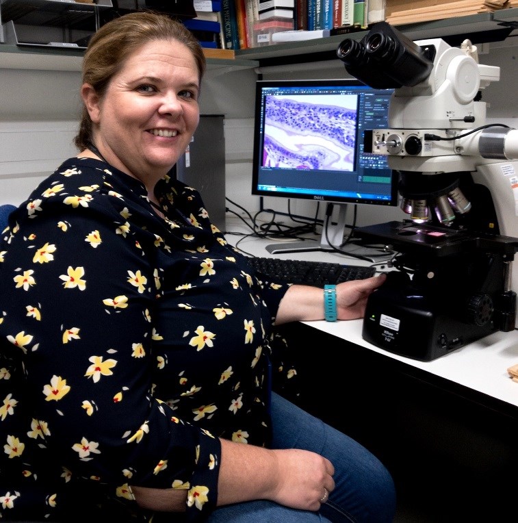
The pathology team uses a range of microscopy techniques to study the anatomy of cells, tissues and organs. The techniques are used to study the health status of aquatic animals, investigating infectious and toxicologic aquatic animal diseases. This includes the diagnosis and description of new and emerging aquatic animal pathogens of concern, the Pathology team is central to the operation of the World Organisation for Animal Health (WOAH) Collaborating Centre for Emerging Aquatic Animal Diseases https://www.cefas.co.uk/icoe/aquatic-animal-health/designations/woah-collaborating-centre-for-emerging-aquatic-animal-disease. The description of novel/emerging pathogens is crucial for disease management in aquaculture systems and hence the Pathology team is also central to the Seafood Hazards and Food from Water Themes, and the International Centre of Excellence for Aquatic Animal Health.
How to work with us
The histology laboratory combines traditional and state-of-the-art technologies for preparation of tissues for histopathological assessment. High throughput systems for embedding, processing, staining and reading of specimens are contained within the ISO 17025 accreditation framework for diagnosis of aquatic animal pathogens. The unit offers a comprehensive array of services for light microscopy including histological special stains, cryotomy, in situ hybridisation and immunohistochemistry. High-resolution digital image capture systems and user-protected data storage ensure rapid and auditable result trails. Using a high throughput digital slide scanner and online digital pathology portal, we also offer a digital slide scanning service, pathology consultancy and training. In addition, molecular pathology techniques are achievable using our Laser capture microdissection (LMD) facility, for in-situ molecular analysis of histological tissue sections e.g. pathogen identification within coinfections and single cells.
The ability to accurately diagnose previously unrecognised disease conditions is dependent on familiarity with a wide range of pathogens, or potential pathogens, infecting hosts from a variety of sources. The Registry of Aquatic Pathology (RAP) is a unique reference collection of pathological conditions in aquatic animal hosts and includes examples of disease conditions (viral, bacterial, fungal, parasitic, environmental) from cultured and wild fish, bivalve molluscs and crustacea from freshwater and marine environments around the world. New examples are continually being added, and records are updated to ensure the information provided is still accurate.
Transmission Electron Microscope (TEM) allows for ultrastructural examination of cells for changes caused by exposure to pathogens and environmental stressors. Further it provides a key resource for the identification of novel pathogens (from viruses to parasites), particularly prior to the development of molecular tools for their diagnosis. As such, TEM is the only means of directly visualising some pathogens such as viruses.
Histology and Accreditation = Matthew Green (matthew.green@cefas.co.uk)
Digital Pathology and Registry of Aquatic Pathology (RAP) = John Bignell (john.bignell@cefas.co.uk)
Transmission Electron Microscopy = Stuart Ross (stuart.ross@cefas.co.uk)
Kelly Bateman