View Record
857
Cancer pagurus
Crab, Edible Or Brown
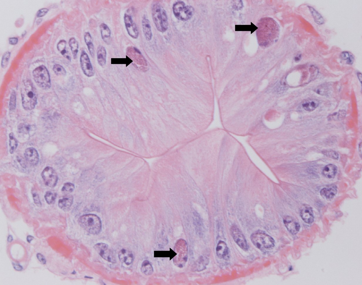
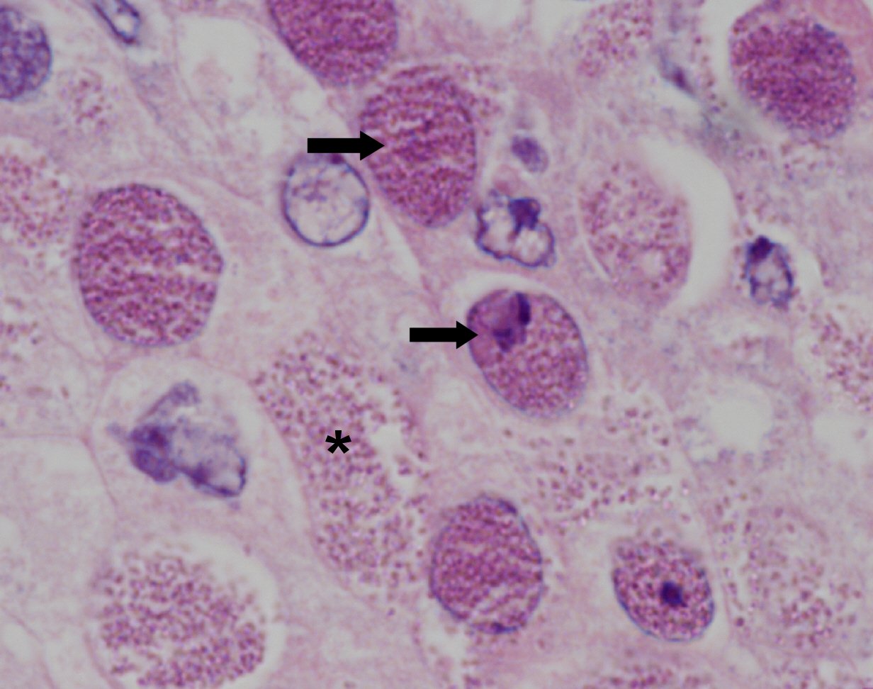
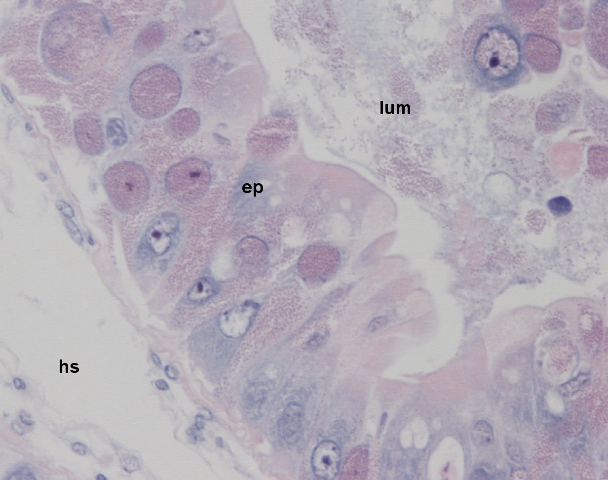
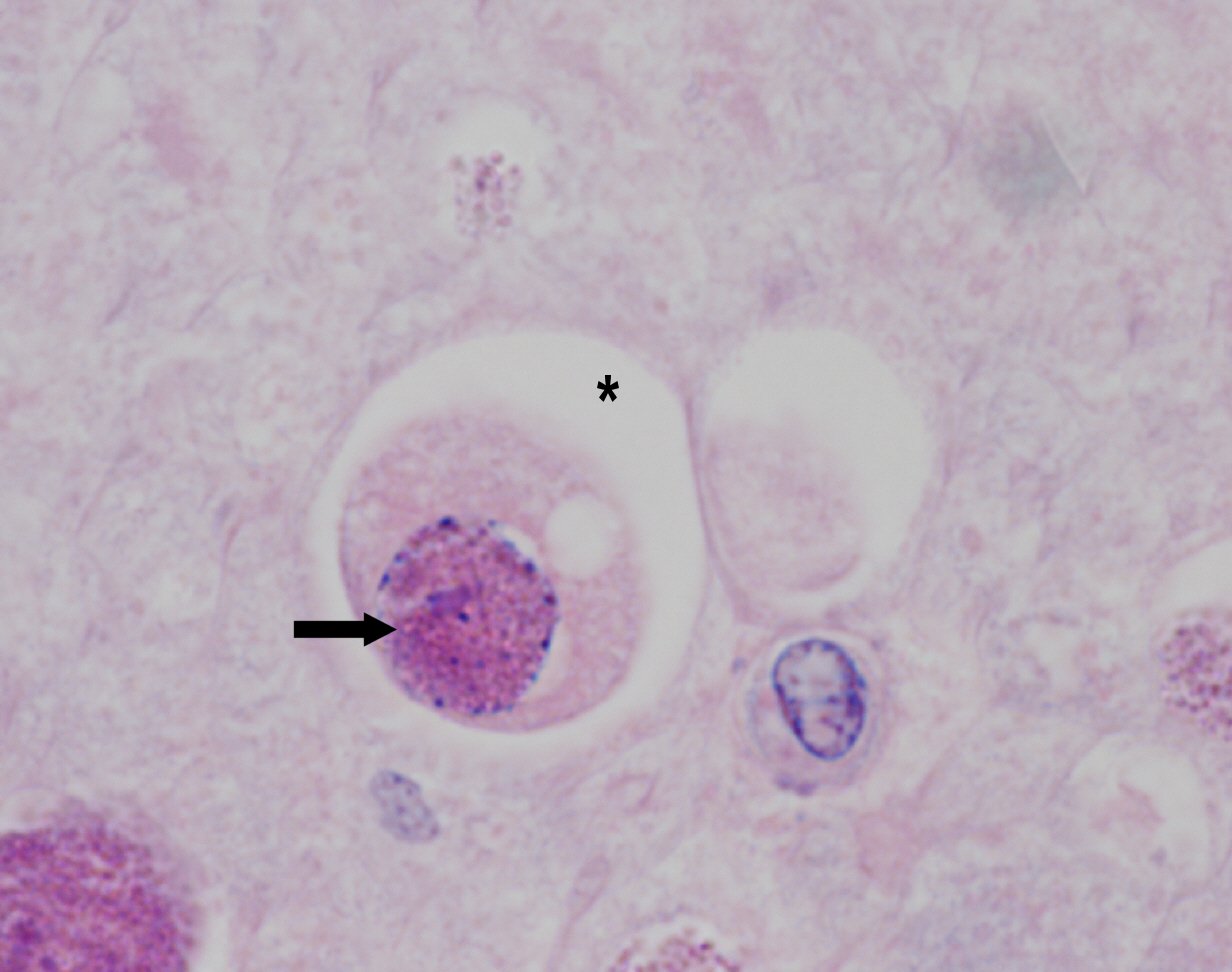
Intranuclear microsporidian
Hepatopancreatic epithelial cells
Enterospora canceri
Enterospora canceri
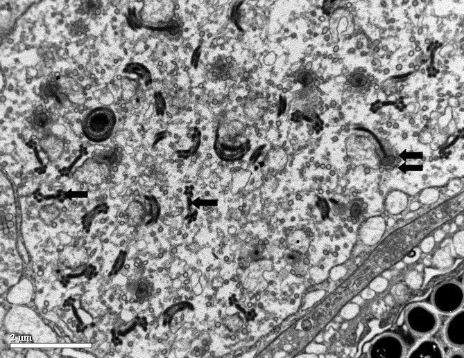
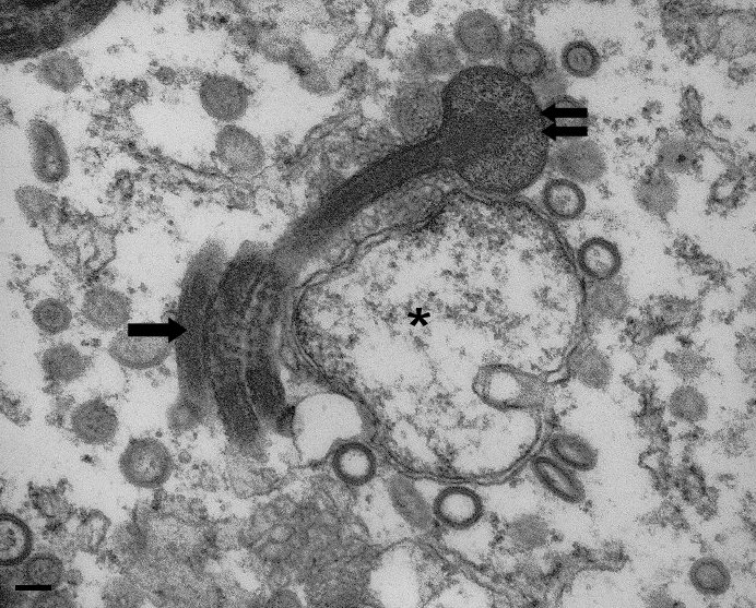
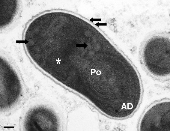
Spores measure 1.3 ± 0.02 x 0.1± 0.01µm, ovoid, central nucleus within sporoplasm, 4-5 turns of polar filament, bell-like polaroplast and obliquely positioned posterior vacuole. Infect nuclei of hepatopancreatic epithelial cells
Stentiford, G.D., Bateman, K.S., Longshaw, M., Feist, S.W. (2007) Enterospora canceri n. gen., n. sp., intranuclear within the hepatopancreatocytes of the European edible crab Cancer pagurus. Diseases of Aquatic Organisms, 75, 61-72.
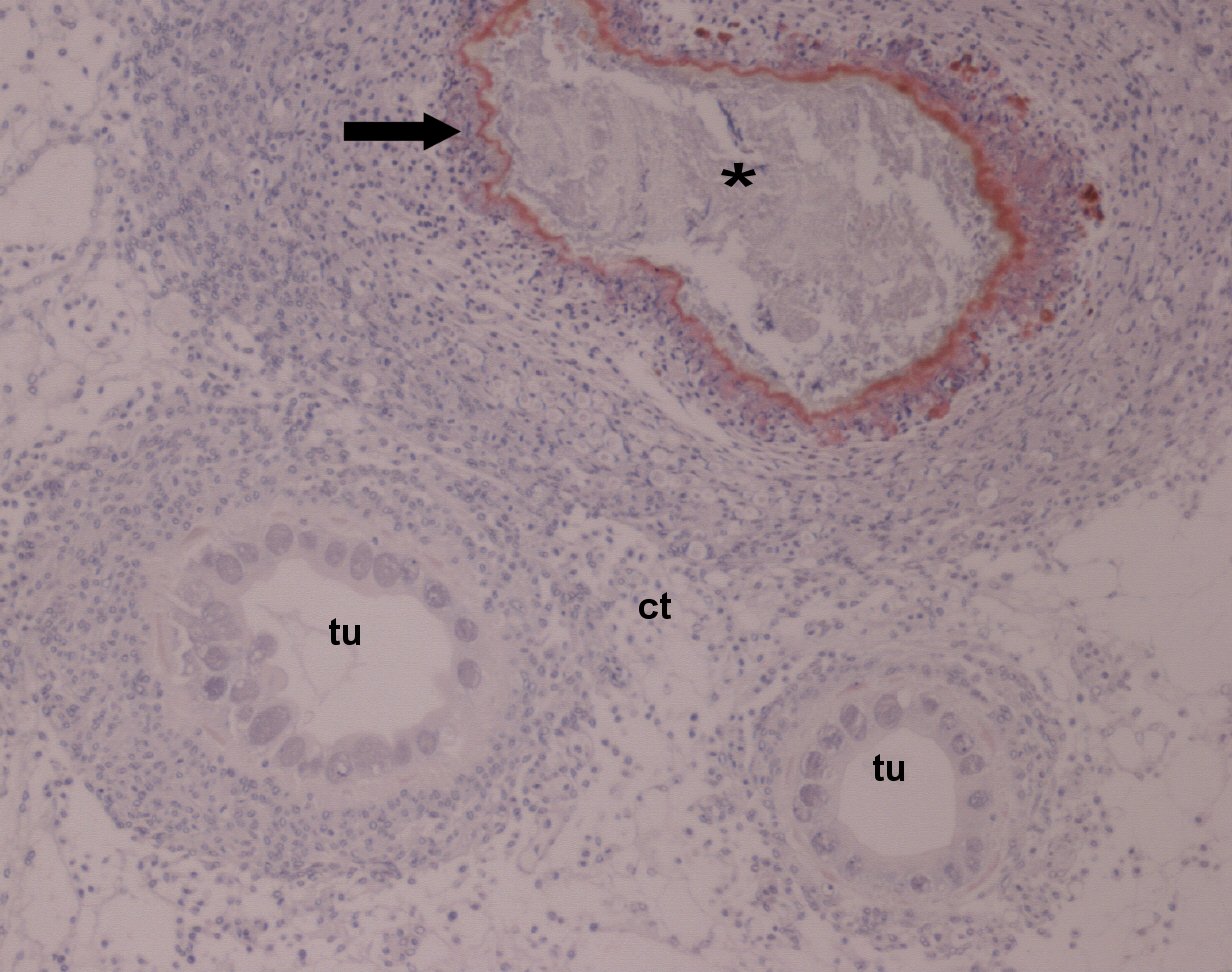
Mortalities unknown as newly emerged disease. Infected crabs did not appear to display external symptoms of disease, white focal patches were occasionally observed on the hepatopancreas.
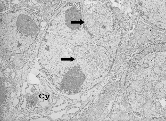
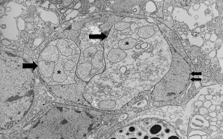
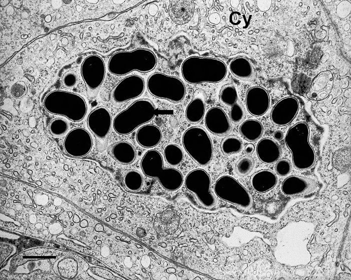
Affected cells included the R (reserve), F (fibrillar) and E (embryonic)-cells of the hepatopancreatic tubules, with B (blister)-cells apparently unaffected. Displacement of the basophilic nucleolus and margination of the chromatin occurred. Parasite stages appear as eosinophilic granular bodies within hypertrophic nuclei. In addition to changes within the nucleus, some epithelial cells may contain cytoplasmic infections associated with degeneration of the tubule epithelia occurred. Parasites and sloughed epithelial cells can be present within the tubule lumen. In heavily affected specimens, the majority of nuclei contain eosinophilic inclusions and degeneration of the hepatopancreatic tubules is evident. Cells often appeared to separate from their neighbours and a host response may be seen with affected tubules becoming encapsulated with mixed populations of hyalinocytes and granulocytes. Necrotic tubules, demarcated with melanin deposition are frequently observed. No microsporidian stages were detected in other host organs.
English Channel
U.K
K.S.Bateman
3/30/2004