View Record
869
Eriocheir sinensis
Crab, Chinese Mitten
Microsporidian
Hepatopancreatic tubules
Endoreticulatus eriocheir
Endoreticulatus eriocheir
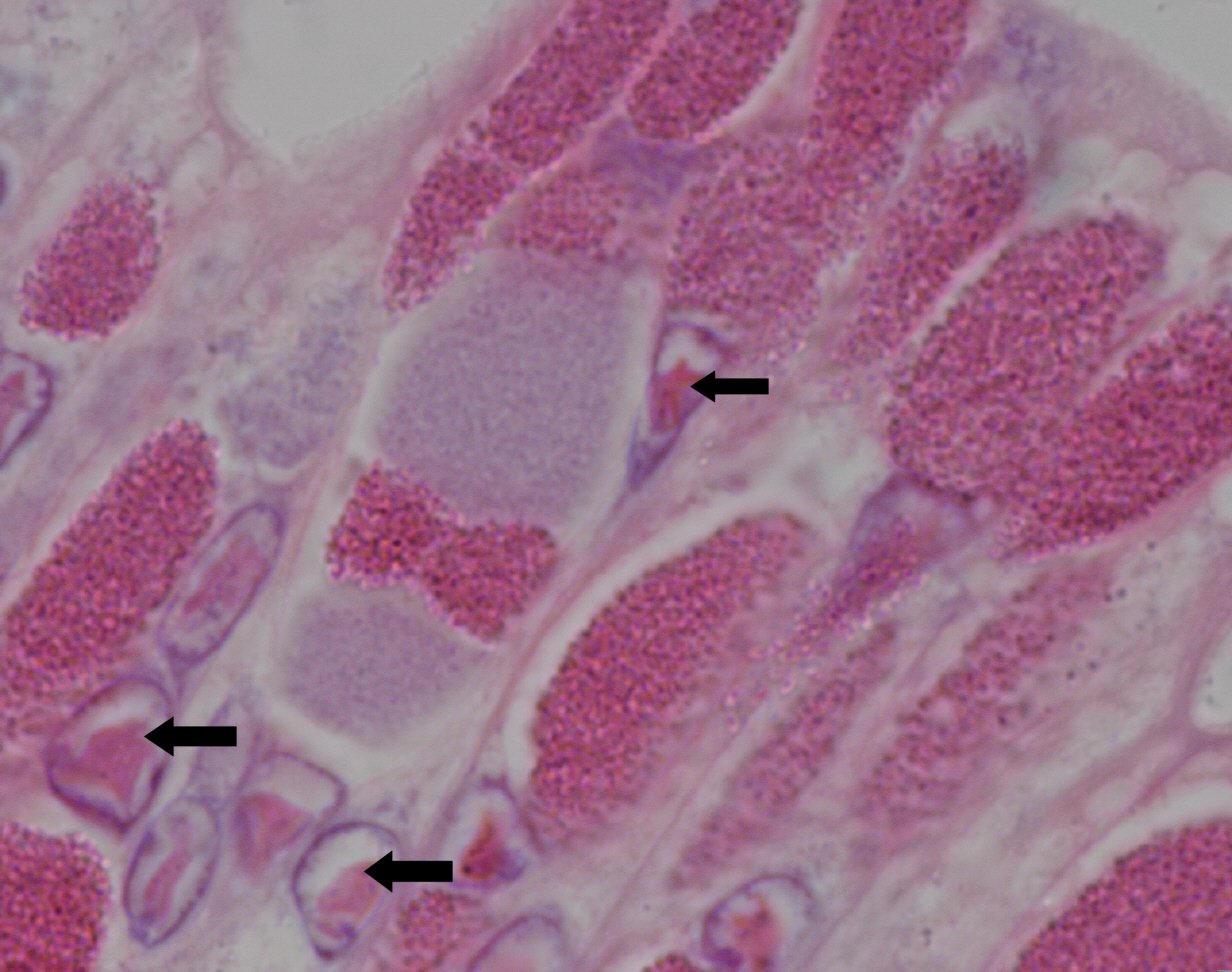
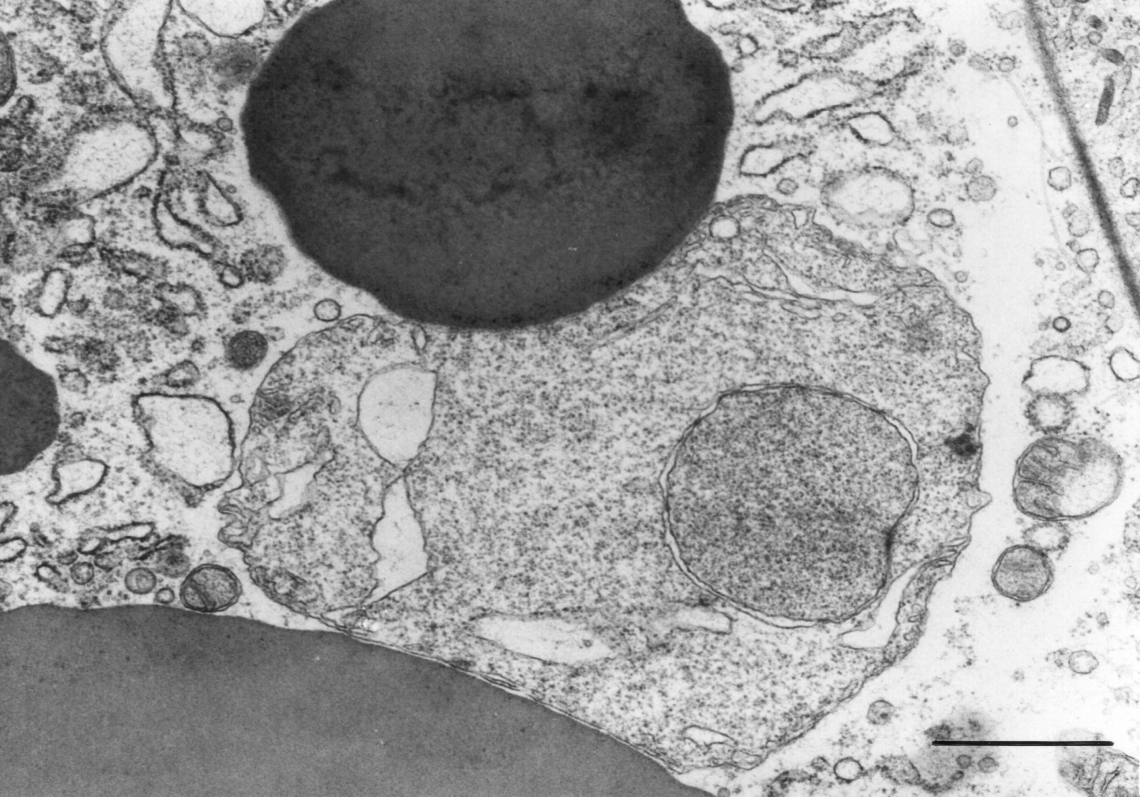
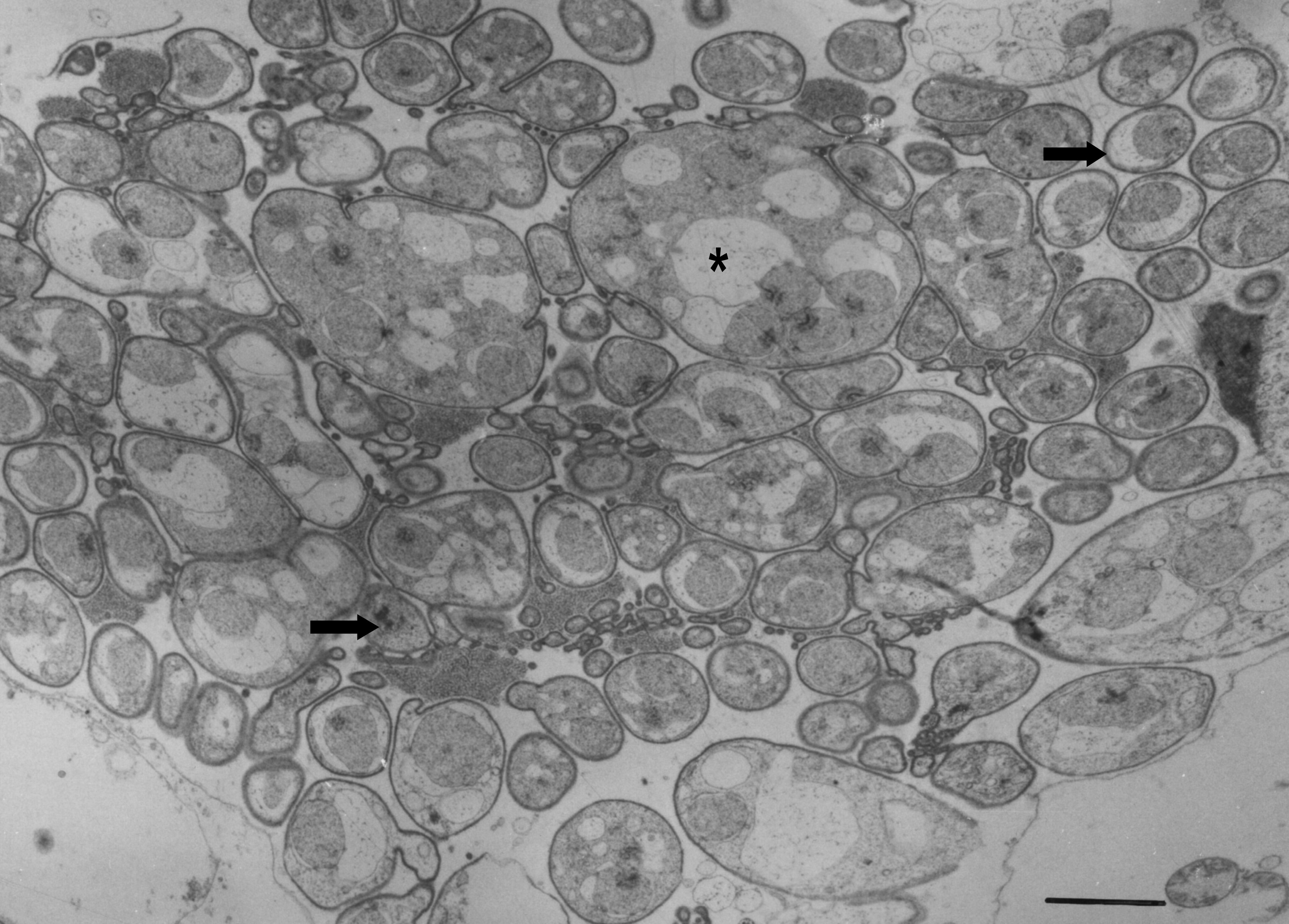
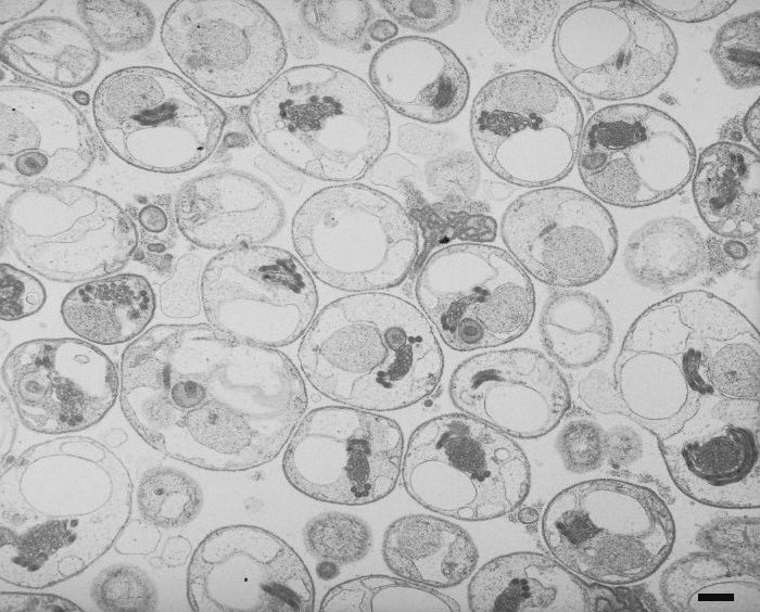
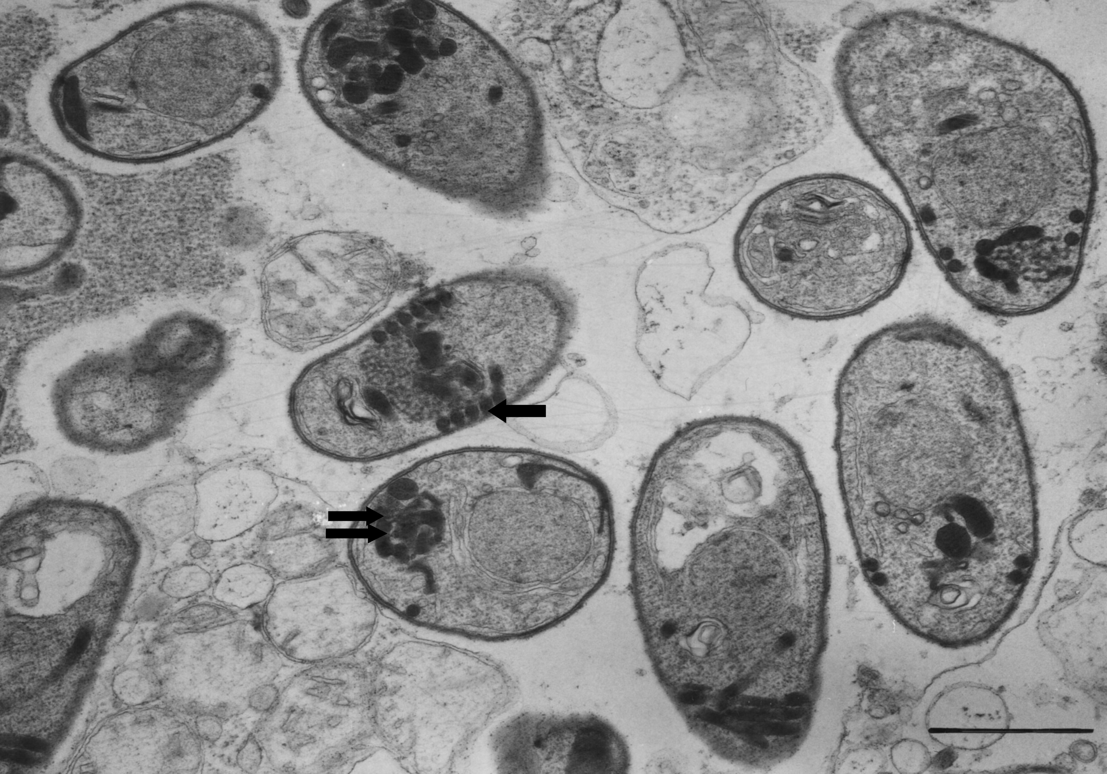
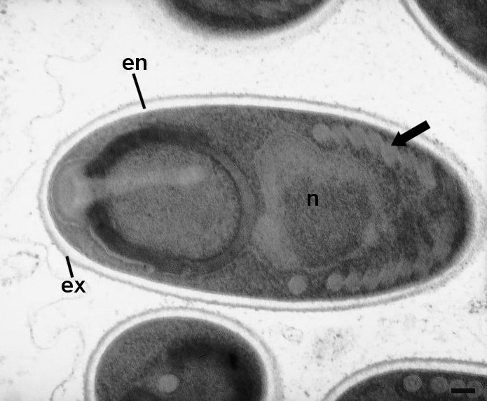
Ellipsoidal, spores measure 1.7µm x 1.0µm, central nucleus within sporoplasm, 7 turns of polar filament, bell-like polaroplast and obliquely positioned posterior vacuole
Wang, W. and Chen, J., (2007) Ultrastructural study on a novel microsporidian, Endoreticulatus eriocheir sp. Nov (Microsporidia, Encephalitozoonidae), parasite of Chinese mitten crab, Eriocheir sinensis (Crustacea, Decapoda). J Invertebr Pathol, 94: 77-83
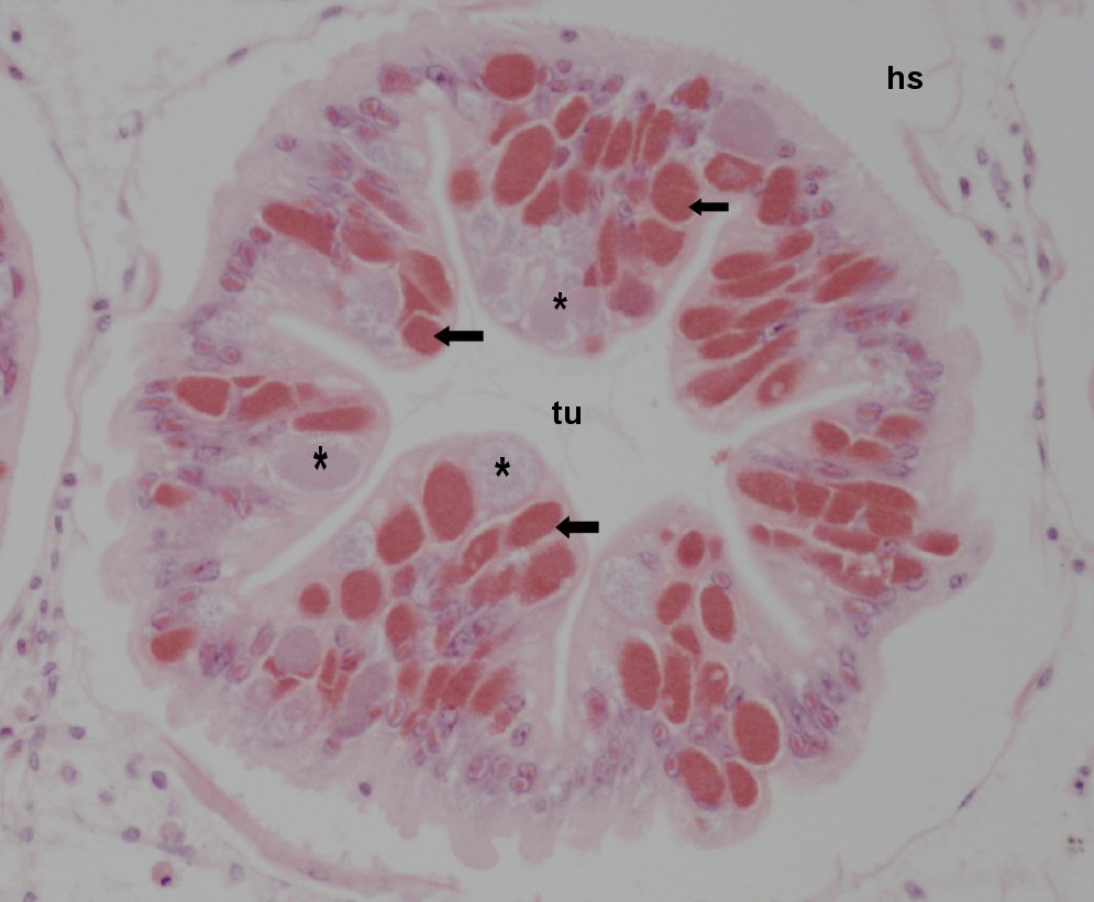
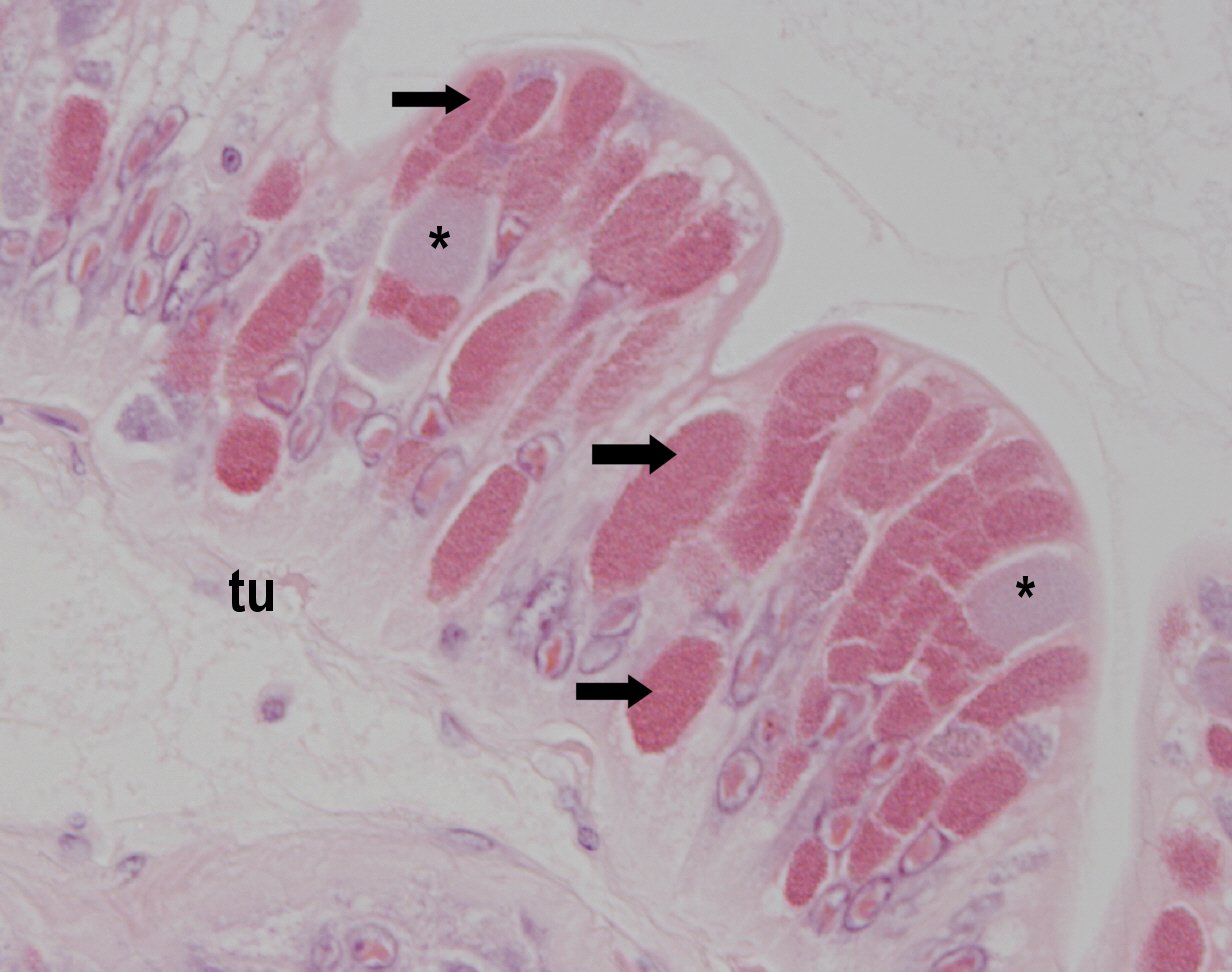
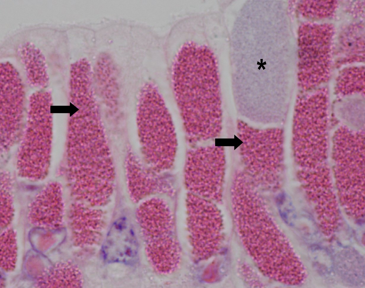
Mortalities unknown as newly emerged disease. Infected crabs did not appear to display external symptoms of disease, white focal patches were occasionally observed on the hepatopancreas.
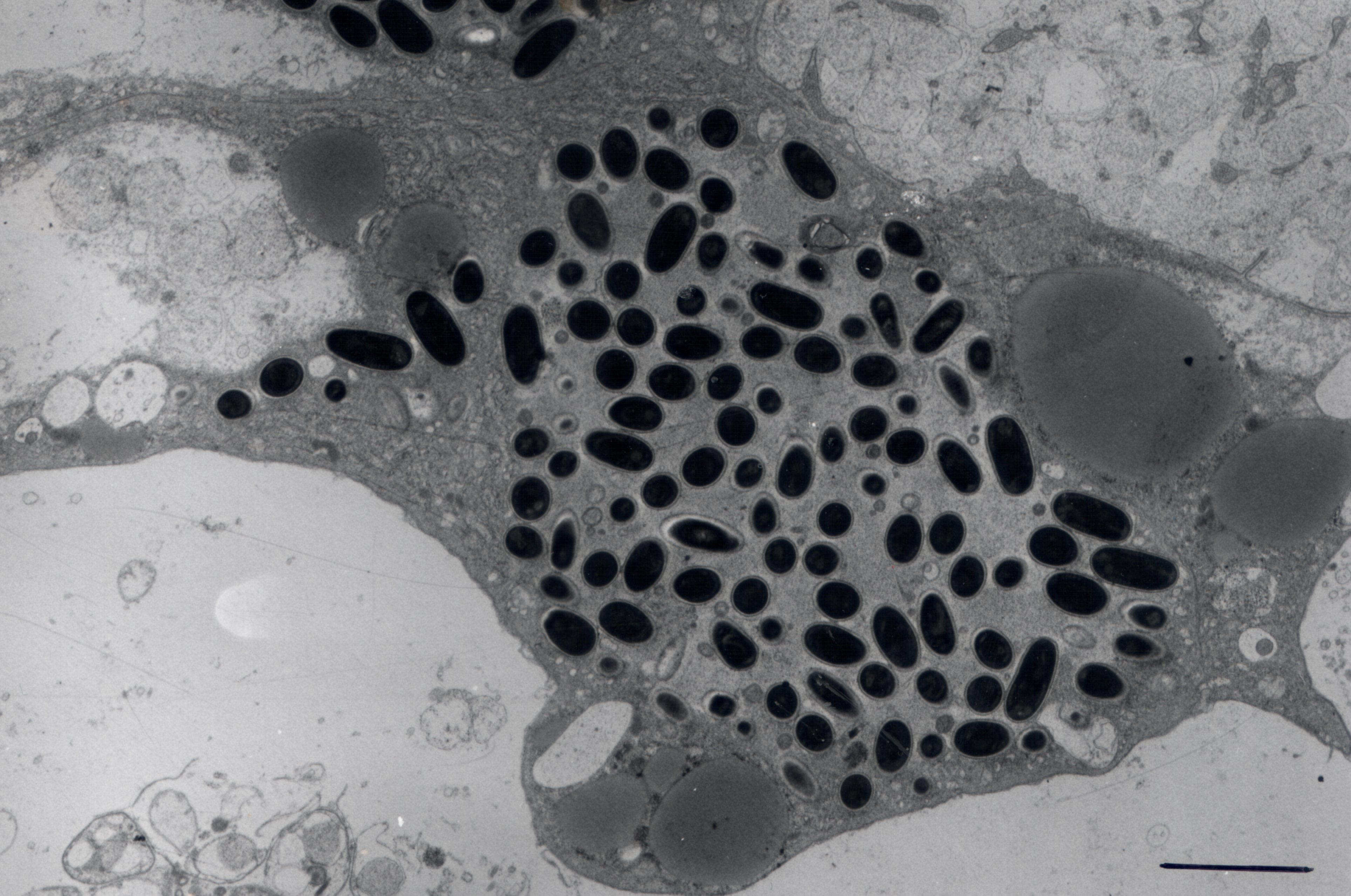
Epithelial cells within infected hepatopancreatic tubules contain single basophilic or eosinophilic packets, some of which may contain multiple packets. Staining characteristics relate to different development stages of the parasite, eosinophillic staining corresponding to meront stages and basophilic staining relating to sporont stages. In severely affected tubules, the majority to all of the epithelial cells are infected. Infection is confined to the epithelial cells of the hepatopancreas, with no other organ seemingly infected. In severe cases hepatopancreatic tubules become disrupted, liberated parasite spores can be observed within the lumen of affected tubules and in the haemolymph surrounding the tubules. Host inflammatory reactions can be seen in response to tubule debris and liberated spores.
River Thames
London
U.K
K.S.Bateman
11/5/2003